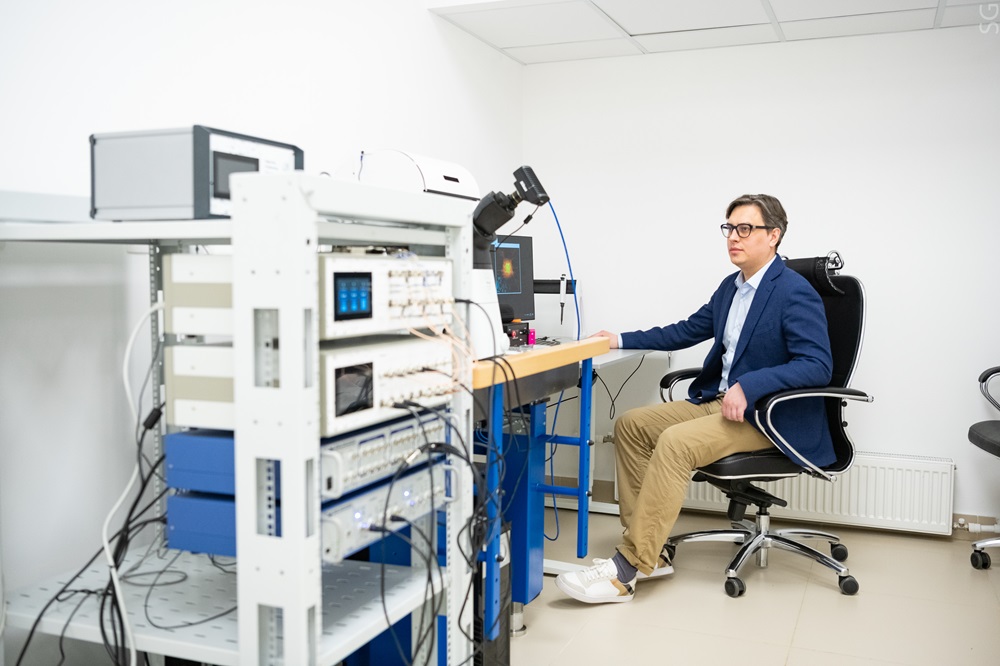
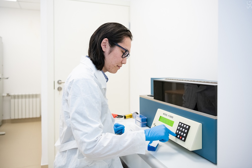
The research group of NUST MISIS, along with colleagues from the Russian Academy of Sciences’ Institute of Molecular Biology, demonstrated the efficiency of a fundamentally new method for analyzing neuronal cells. Using the only scanning ion-conducting microscope in Russia with a confocal module, scientists have revealed that Alzheimer’s disease causes neuronal cells to become mechanically stiffer because amyloid aggregates formation on their surface.. Novel therapeutic agents for Alzheimer’s disease treatment are currently being examined using this innovative scientific equipment.
Researchers showed that the properties of neuronal cells — size, density, and flexibility — can indicate a risk of ischemia, trauma, Parkinson’s illness, or Alzheimer’s disease. Alzheimer’s disease is caused by the aggregation of β-amyloid oligomers (Aβ), which disrupts neuronal function. Scanning ion-conductance microscopy is the most efficient technique for studying the formation of Aβ aggregates on neuron surfaces.
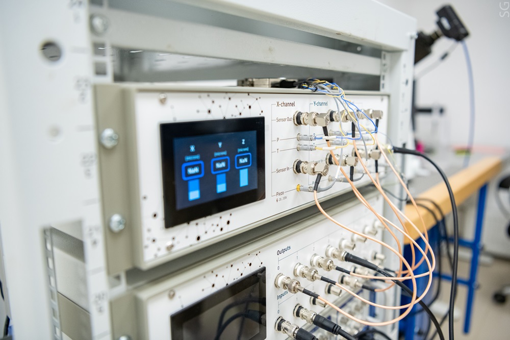
“The scanning ion-conducting microscope with a confocal module is the only unique scientific equipment in Russia. We are proud that it is located at the National University of Science and Technology MISIS. It allows the study of live cells under conditions close to physiological ones, with minimal impact on the neuron sample,” said Alevtina Chernikova, Rector of NUST MISIS.
Other methods of studying neurons are challenging because they can damage the cells. Atomic force microscopy is the nearest analogue to this problem, which has provided the majority of the results. However, one known disadvantage is the considerable impact on the samples.
In the new study, the structure of Aβ was characterized, and the cell mechanical stiffness was determined. Knowing the magnitude of deformation allows determining the mechanical stiffness of cells. This is essential to studying the cytoskeleton, which is the structure that gives a cell its shape and allows it to resist external stresses. The cytoskeleton and mechanical properties of neuronal cells play key roles in signal transmission in the central nervous system. When these are disrupted, cognitive issues can develop.
“We have established that cell stiffness increases after the formation of Aβ on the surface of the cell membrane. Upon closer examination, it can be seen that a pore forms on the neuron’s surface. Aβ is embedded in it, disrupting the normal ion homeostasis of the cell,” noted Vasily Kolmogorov, an engineer of the Research Laboratory of Biophysics at NUST MISIS.
Moreover, platinum nanoelectrodes were used to study the effect of Aβ on reactive oxygen species (ROS). Aβ on the cell surface considerably increases ROS production. It indicates that the presence of amyloid peptides on the cell membrane surface affects the level of oxidative stress, which may lead to cell death. The detailed results of the study are described in one of the highranked scientific journals, ACS Publications (Q1).
“This work can serve as a basis for further research into determining a substance’s ability to exert toxic effects on cells. The developed methodology provides an opportunity to test the latest drugs aimed at therapy for neurodegenerative diseases at the cellular level,” said Alexander Erofeev, PhD., Head of the Research Laboratory of Biophysics at NUST MISIS.
The work is supported by the Ministry of Science and Higher Education of the Russian Federation. The research theme is aimed at solving tasks within the framework of the “Development of large-scale scientific and scientific-technological projects in priority research areas” project as part of the “Science and Universities” national project.