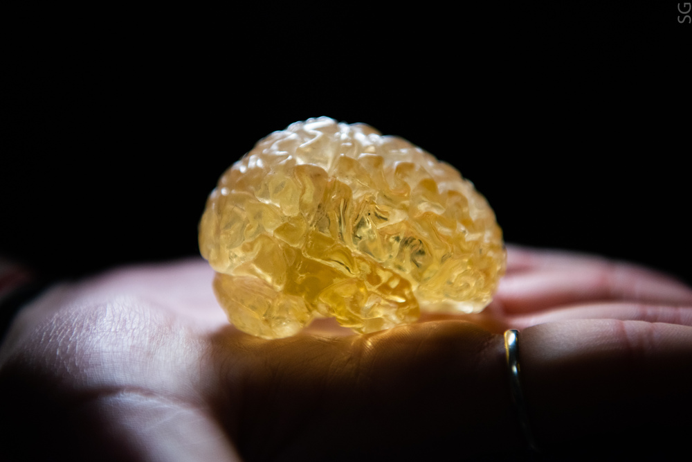
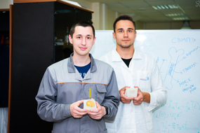
A team of NUST MISIS students developed a “phantom” of the human brain, i.e. a hydrogel model with structural and mechanical similarity to a real organ. “Fantomozg” (en: Phantombrain) will allow students and neurosurgeons to study the pathological anatomy of tissues and train in complicated surgical manipulations.
Neurosurgery is a very complicated process that requires full understanding of the brain structure, the nature and form of pathology, and the acceptable limits of intervention. Even neurosurgeons with many years of experience can face difficulties with a rare clinical case, whether it is a difficult-to-reach tumor location, a massive stroke, or a particularly large hematoma.
Today, surgeons and students have three main ways to study the structure of organs: through 3D simulation in VR, on cadavers (i.e., human bodies), and using real-size phantom models of organs. Each method has its drawbacks. For example, VR technologies are still very expensive, and do not allow the operator to fully experience the operation process physically. The use of cadavers is inhumane, in addition, dead tissue decomposes and loses its original structure anyway. Phantoms are the best option, but today they are made of silicone, which is very different from human tissue in terms of mechanical characteristics.
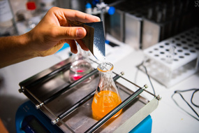
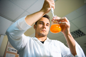
NUST MISIS students suggested an alternative version of manufacturing a human brain phantom, i.e. from hydrogel, based on the patient’s computer and magnetic resonance imaging data. The first stage is 3D reconstruction followed by printing of the polymer negative. Then, based on the negative, a silicone mold is made, into which a hydrogel (polyvinyl alcohol and agarose) is poured. The resulting billet is placed first in the freezer, and then in the refrigerator. The total production time of such a phantom is about 30 hours.
The test sample was a reduced copy (scale 1:4); mechanical tests showed that the ultimate strength of the phantom almost corresponds to the hemispheres of the human brain, 87 kPa vs 100 kPa. Next, the team will work on structural simulations of other parts of the brain (cerebellum, midbrain, medulla oblongata, hindbrain), as well as increasing the model to the size of 1:1. In addition, it is planned to add to the phantom simulation of vessels and pathological changes: tumors, blood clots, plaques.
The work is being carried out at NUST MISIS Center for Composite Materials, Skoltech University with the support of “Art, Science and Sport” charity foundation.