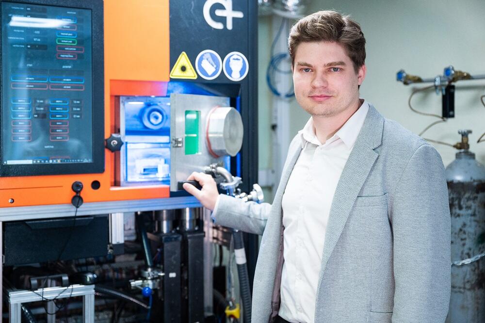
 Scientists find a way to “program” metal behavior during 3D printing
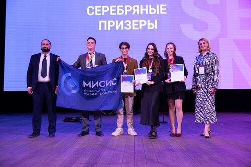 NUST MISIS Students Become Prize Winners of the 14th International Engineering Championship CASE-IN
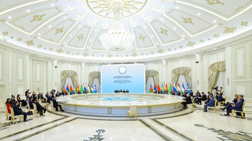 MISIS University Participated in the CIS Council of Heads of Government Meeting
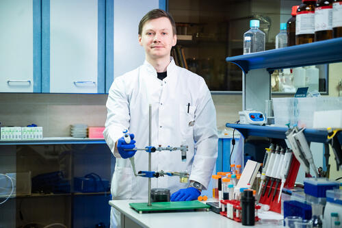 Scientists at NUST MISIS Taught Magnetic Nanoparticles to Remove Dyes from Water