Russian researchers have improved an anti-scatter grid for
During an
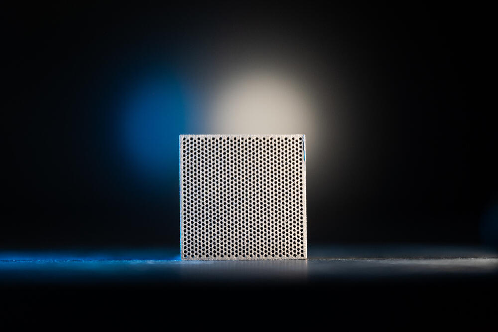
Researchers at NUST MISIS, the Institute for Nuclear Research of the Russian Academy of Sciences, the Kurchatov Institute Research Centre, and Moscow Polytechnic University have proposed using 3D printing to manufacture tungsten grids with a new design featuring converging channels. Unlike conventional structures with parallel partitions, the channels in the new filter are aligned with the direction of the
“We successfully produced a thin-walled grid structure using selective laser melting of micron-sized tungsten powder. This approach makes it possible to significantly reduce the thickness of structural elements and create the geometry required for the beam of a specific
X-ray system. We then tested the grid under conditions close to those of real diagnostic imaging.X-ray images were taken using a model of the human pelvic region, with special test markers placed at points that needed to be distinguished in the image. Without the filter, the image consisted largely of background noise, and the shadows of the markers were barely visible. With the grid in place, however, they could be clearly seen,” Stanislav Chernyshikhin, Head of the Additive Manufacturing Laboratory at MISIS University.
To assess the tungsten grid’s performance under more challenging conditions, the researchers placed a layer of acrylic glass in front of the pelvic model to simulate human tissue. Without the grid, the test markers were almost completely lost in the noise, whereas with the filter, they remained clearly distinguishable. Detailed results of the study were published in the journal Bulletin of the Lebedev Physics Institute.
“Another challenge was ensuring that the filter itself did not interfere with image acquisition, since even thin partitions leave their own imprint on the image. We demonstrated that this shadow can be removed through digital processing using the fast Fourier transform without any loss of diagnostic information. The same approach is used with conventional lead filters,” Igor Dyachkov, an engineer at the MegaScience Infrastructure Cooperation and Partnership Center at MISIS University.
According to the researchers, the benefits of a thinner grid will be particularly noticeable when examining dense areas of the body, where scattered radiation causes the greatest image distortion. These are precisely the scenarios that pose the greatest challenges in clinical practice.