A Russian-German research team has come up with a new technique for magnetic resonance imaging of cancer cells. The study, published in Pharmaceutics, shows that heterologous expression of encapsulin systems from Quasibacillus thermotolerans with functional cargo proteins and iron transporter leads to increased contrast in MRI imaging of mammalian tumor cells.
Many advances in cancer treatment would come from a better understanding of tumor biology, particularly the elucidation of carcinogenesis mechanisms.
Currently, the primary method of live-cell imaging is direct labeling of cells with a probe or contrast agent before transplantation. However, any synthetic contrast agent for cell labeling has a critical drawback—it dilutes as the cells divide, which leads to loss of the signal after several cycles of divisions. In contrast, genetically encoded reporters propagate to daughter cells with each cell division. Moreover, because genetically encoded reporters rely on essential cellular processes, their signal is selective for viable cells.
The most commonly studied genetically encoded labels use an optical signal generated by either bioluminescent or fluorescent reporter protein. Although these methods have very high sensitivity, their use is limited by light scattering in biological tissues.
MRI has the advantage of deep tissue penetration with relatively high spatial resolution. Ferritin, a blood protein that contains iron, is the most studied genetically encoded agent so far. Nevertheless, ferritin’s MRI performance is severely limited by its weak magnetic properties and highly conservative structure. The latter excludes significant improvement in ferritin relaxivity by bioengineering.
“One of the most promising approaches is based on the heterologous expression of bacterial protein nanocompartments—encapsulins— in mammalian cells. Encapsulins, which are bacterial protein nanocompartments, can serve as genetically controlled labels for multimodal detection of cells. Such nanocompartments can host various guest molecules inside their lumen. These include, for example, fluorescent proteins or enzymes with ferroxidase activity leading to biomineralization of iron oxide inside the encapsulin nanoshell. Besides, these reporters do not suffer from dilution during cell division,” says Maxim Abakumov, head of the NUST MISIS Biomedical Nanomaterials Laboratory, senior researcher at the Medicinal Nanobiotechnology Department, N.I. Pirogov Russian National Research Medical University.
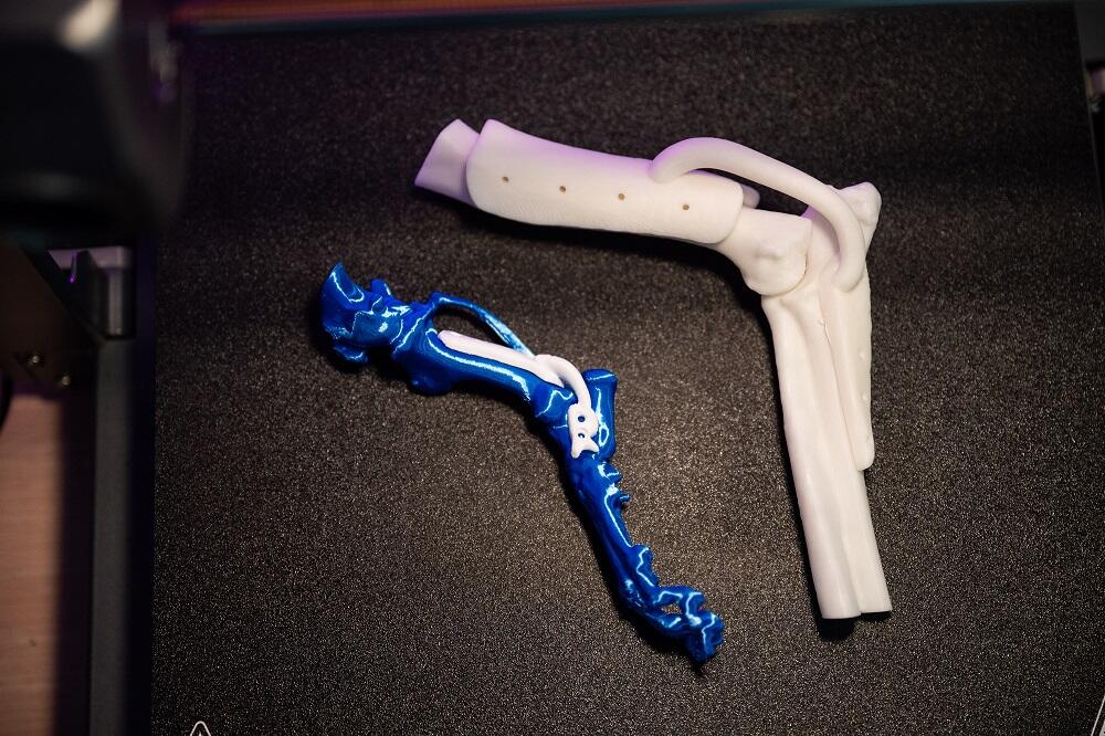
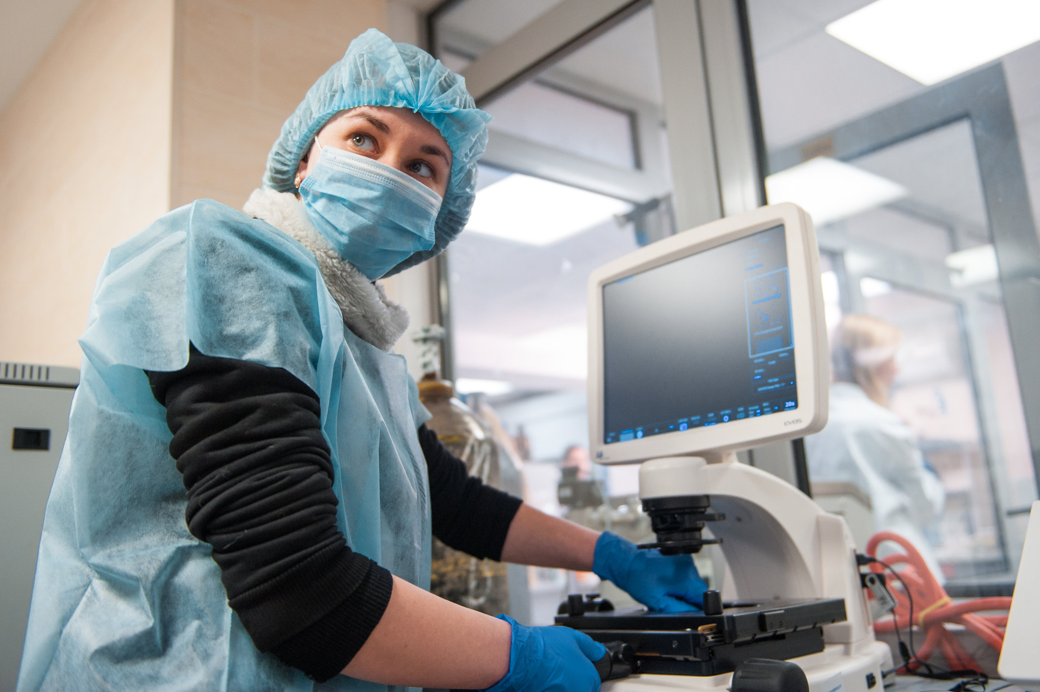
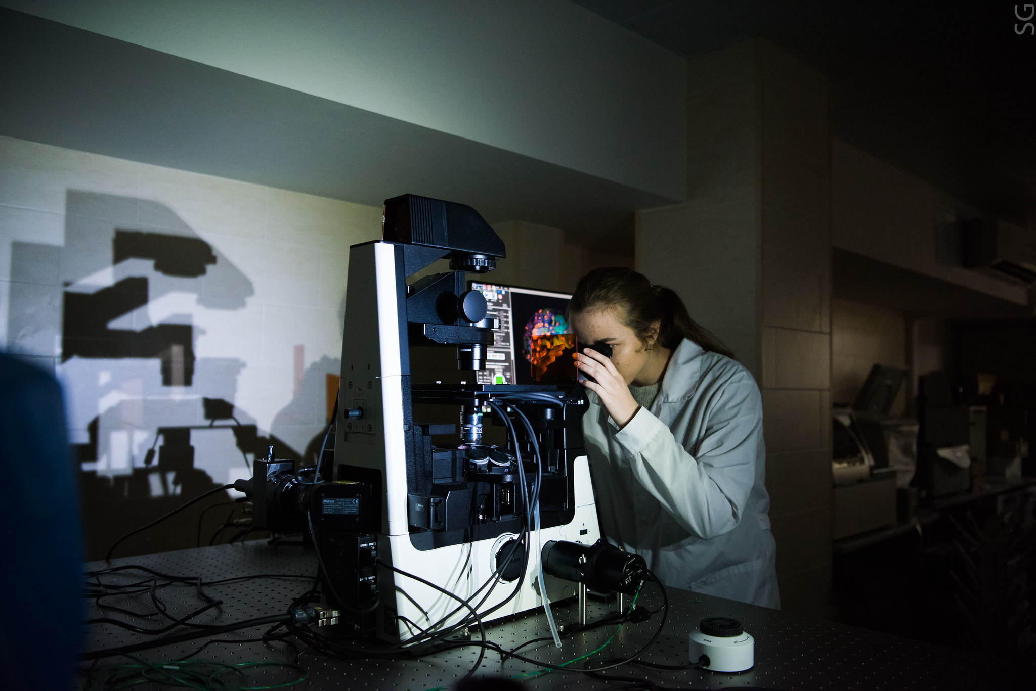
In their experiments, a team of scientists from NUST MISIS, V. Serbsky National Medical, N.I. Pirogov Russian National Research Medical University, Technical University of Munich, Helmholtz Center Munich have implemented, for the first time, heterologous expression of encapsulin systems from Quasibacillus thermotolerans using a fluorescent reporter protein and ferroxidase in human hepatocellular carcinoma cells. Gene expression is the process by which information from a gene is used in the synthesis of a functional gene product that enables it to produce end products, protein or non-coding RNA, and ultimately affect a phenotype, as the final effect. The researchers loaded the nanoshell with the natural ferroxidase cargo from Q. thermotolerans and a synthetic fluorescent cargo protein derived from mScarlet-I. The successful expression of self-assembled encapsulin nanocompartments with functional cargo proteins was then confirmed by fluorescence microscopy and transmission electron microscopy. Also, coexpression of encapsulin nanoshells, ferroxidase cargo, and iron transporter led to an increase in contrast in magnetic resonance imaging of cancer cells. The encapsulin cargo system from Q. thermotolerans may be suitable for multimodal imaging of cancer cells, the researhers believe.