A Russian-Canadian research has found that stem cell transplantation may help restore brain function in stroke patients. The study was published in Frontiers in Neuroscience.
Ischemic stroke, also known as brain ischemia and cerebral ischemia, is caused by a blockage in an artery that supplies blood to the brain. Ischemic stroke is one of the prevailing causes of disability and death throughout the world, with over 450 000 cases registered annually in Russia alone. The immediate therapeutic objective for acute ischemic stroke patients is prompt and efficient restoration of blood flow to ischemic brain tissue. This can be achieved by administering a clot -busting drug in a process called thrombolysis or mechanical thrombectomy, a type of a minimally-invasive procedure in which an interventional radiologist uses specialized equipment to remove a clot from a patient’s artery. Both methods have limitations and are effective only if applied during a narrow “therapeutic window” after stroke onset, which is
In their experiments, the scientists from the Pirogov Russian National Research Medical University, NUST MISIS and New World Laboratories used reprogrammed human neural precursor cells (NPC) and mesenchymal stem cells (MSC). NPCs generate neurons and glial cells and thus are obvious substitutes for the recipient’s own ones eradicated or impaired by stroke. Moreover, both neural and mesenchymal stem cells produce different substances that promote brain tissue regeneration.
MSCs can readily be obtained from different sources, including the umbilical cord, cord blood, adipose tissue, and bone marrow. However, isolation of native human NPC directly from fetal or adult brain is associated with serious technical, legal and ethical problems, alternative methods of their production for preclinical studies and clinical needs have been developed. Currently, direct reprogramming of somatic cells is considered to be the safest way of NPC isolation.
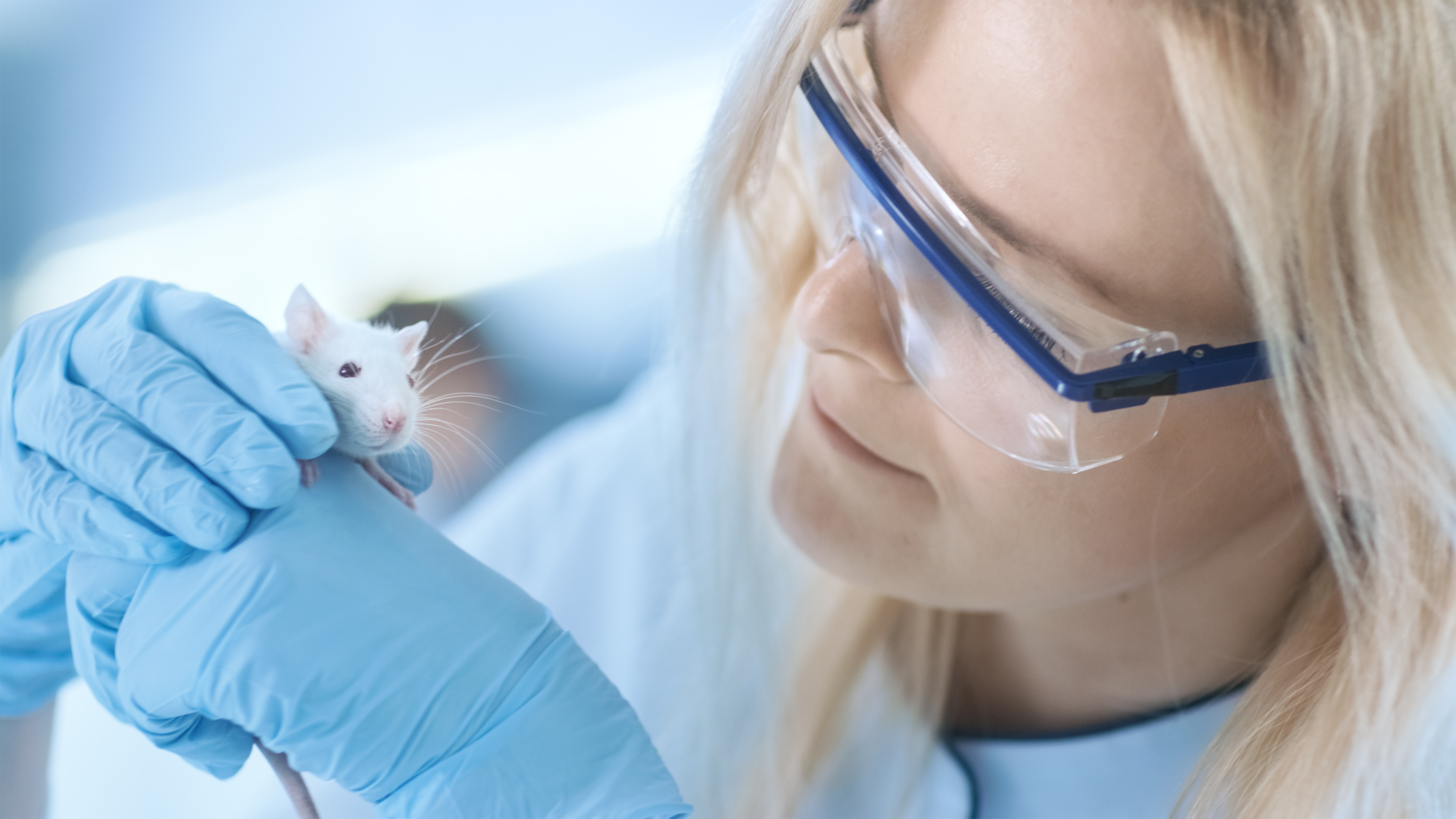
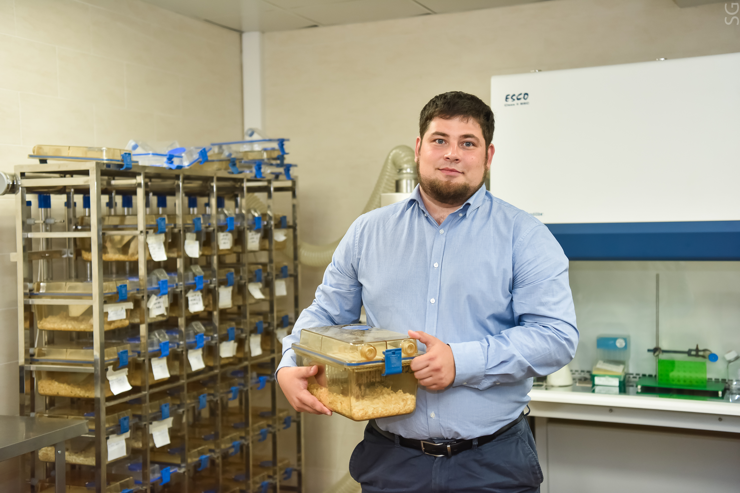
“In this study, the cells were transplanted into rats intra-arterially 24 hours after the transient middle cerebral artery occlusion. Transient middle cerebral artery occlusion in rodents is one of the most widely utilized models in experimental stroke studies. The cells were transplanted intra-arterially to provide targeted delivery to the ischemic lesion and to bypass filtering organs, such as lungs, liver, spleen”, noted the lead author of the study Daria Namestnikova, neurologist, assistant professor at the Department of Fundamental and Clinical Neurology, Pirogov Russian National Research Medical University, senior researcher at the Federal Center of Brain Research and Neurotechnologies of the Federal Medical Biological Agency of the Russian Federation.
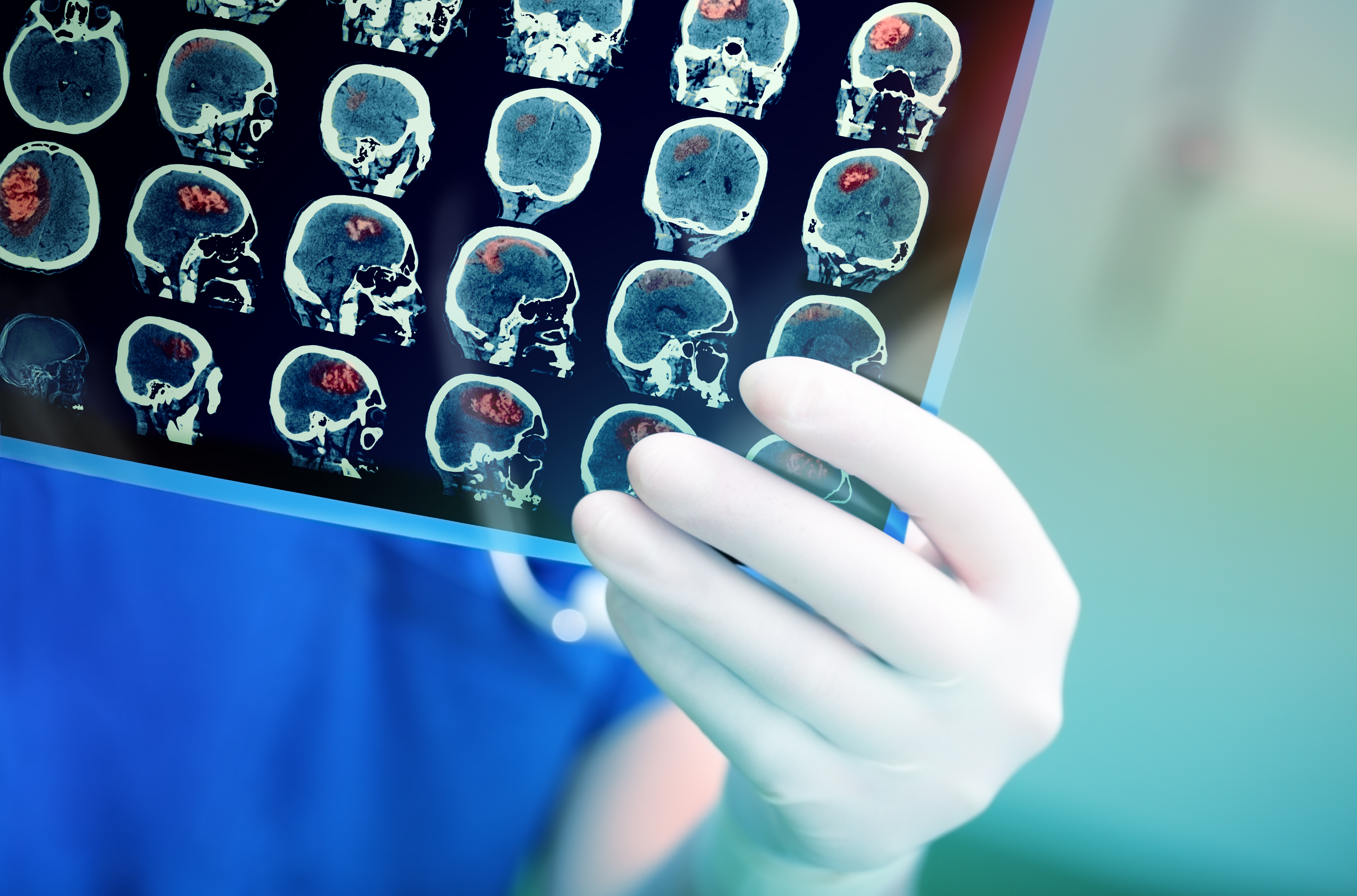
“For precise cell visualization in this study we used real-time MRI starting at the very onset of cell infusion. Immediately after transplantation, cells were observed in the periphery of the infarct zone and in the brain stem, 15 min later small numbers of cells could be discovered deep in the infarct core. Transplanted cells could no longer be detected in the rat brain
48–72 hours after infusion. However, the long-term survival of transplanted cells is not necessary for maintaining continued functional recovery. They may act through an indirect ‘trigger’ mechanism launching a cascade of molecular and cellular events promoting recovery. The positive therapeutical effects were still observed 14 days post-injection”, adds Maxim Abakumov, head of the NUST MISIS Biomedical Nanomaterials Laboratory, senior researcher at the Medicinal Nanobiotechnology Department, N.I. Pirogov Russian National Research Medical University.
Transplanted cells secrete factors (e.g. growth factors) that signal the patient’s cells to change theirs behavior to repair the damaged tissue, in a process called the paracrine effect. In animal stroke models, transplanted NPC most likely influenced brain neural, glial and immune cells and enhanced brain tissue protection and regeneration via secretion of cytokines, growth factors and other biologically active substances.
This treatment method may revolutionize stroke treatment, the researchers believe.