Researchers from NUST MISIS and Lomonosov Moscow State University have developed a method that makes it possible to mechanically stimulate specific regions of living brain cells and observe, in real time, how their properties change. The study revealed that neurons and supporting cells of the nervous system respond differently even to extremely weak mechanical stimuli.
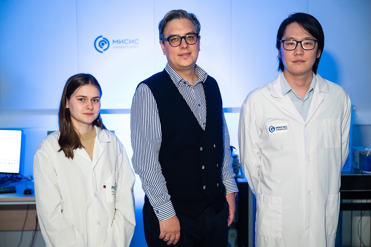
Nerve cells are sensitive not only to chemical and electrical signals but also to mechanical forces arising from injuries and various diseases. However, the mechanisms by which cells detect and process such signals remain poorly understood.
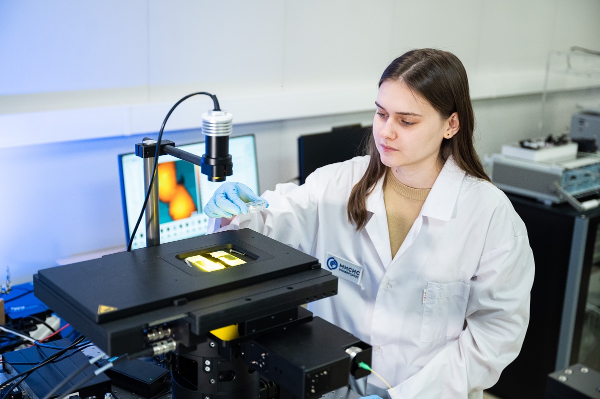
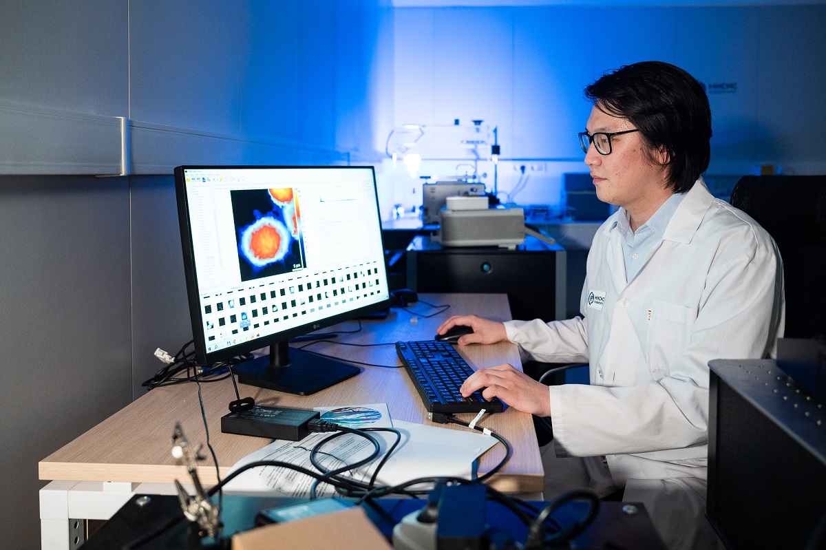
“For several years, a research team at NUST MISIS led by Alexander Erofeev has been developing innovative technologies for medicine. The scientists have created a new approach for studying brain cells based on scanning ion conductance microscopy. Using an ultrafine glass nanopipette, researchers can apply mechanical stimulation to specific regions of a cell with extremely high precision, down to tens of nanometers. The technique does not damage the cell and simultaneously measures changes in its properties in real time. This technology opens up new opportunities for investigating the mechanisms of nervous system function and, in the future, could be used to study the consequences of traumatic brain injuries, neurodegenerative diseases, and the regeneration of nervous tissue after damage,” said Alevtina Chernikova, Rector of NUST MISIS.
The researchers conducted experiments on hippocampal neurons, which is a region of the brain responsible for learning and memory, as well as on astrocytes, the cells that support and protect neurons. They applied localized mechanical stimulation to different parts of the cells and monitored their responses.
“We found that different types of nervous system cells respond to mechanical stimulation in distinct ways. When the cell body of a neuron was stimulated, it became stiffer. This response was associated with the reorganization of the cell’s internal scaffold, or cytoskeleton. In contrast, stimulating neuronal processes produced no such effect. Astrocytes displayed even more complex behavior. Stimulating the central part of the cell caused temporary softening, whereas stimulation of their processes increased stiffness. This suggests that different nervous system cells are able to ‘filter’ physical signals: the metabolic center of neurons is well protected from incidental mechanical disturbances, while the thin processes of astrocytes are highly sensitive to localized mechanical cues, which is critical for sensing external stimuli,” said Alexander Erofeev, Ph.D. in Physics and Mathematics, Head of the Research Laboratory of Biophysics at NUST MISIS.
To determine the cause of these changes, the researchers temporarily blocked the activity of the actin cytoskeleton, one of the key structural components of the cell. After this intervention, the mechanical responses almost completely disappeared. The findings confirmed that cytoskeletal reorganization plays a central role in the cellular response to mechanical stimulation. The results have been published in the journal ChemBioChem (Q1).
“The proposed method not only enables the application of precisely controlled mechanical forces to individual regions of a living cell but also allows researchers to monitor changes in its properties in real time,” said Vasily Kolmogorov, Engineer at the Research Laboratory of Biophysics at NUST MISIS.
The study was supported by a grant from the Russian Science Foundation (Project No.