Russian researchers have found that changes in the “glow” of pigment in retinal cells can be used to detect damage at very early stages, long before serious vision impairment occurs. The scientists also showed how antioxidants slow these processes by reducing the levels of toxic products of photodamage.
Retinal diseases, including age-related macular degeneration, are often diagnosed at late stages, when vision can no longer be restored. One reason is the limitations of existing diagnostic methods: they detect structural changes but miss early functional disturbances in cells.
“Researchers at NUST MISIS have been engaged for several years in developing innovative technologies that in the future will simplify diagnosis and treatment of various diseases. The diagnostic method for retinal pathologies developed at the university based on the ‘glow’ of cells will become an important tool for detecting diseases and assessing the effectiveness of ongoing therapy,” said Rector of NUST MISIS Alevtina Chernikova.
Scientists from MISIS University, Lomonosov Moscow State University, Moscow State Pedagogical University, Moscow Institute of Physics and Technology, and the Shemyakin and Ovchinnikov Institute of Bioorganic Chemistry studied lipofuscin, which is a pigment that accumulates with age in retinal pigment epithelial cells. It can luminesce under light exposure, and its properties can be used to assess the condition of the eye. A key feature of lipofuscin is its phototoxicity: when irradiated with visible light, it can generate reactive oxygen species and toxic oxidation products that cause pronounced oxidative stress. Studying these processes is important for understanding mechanisms of retinal damage and diagnosing age-related degenerative changes. A major role here is played by lipofuscin’s autofluorescence. Measuring its “glow” parameters is an important tool for early diagnosis of eye diseases.
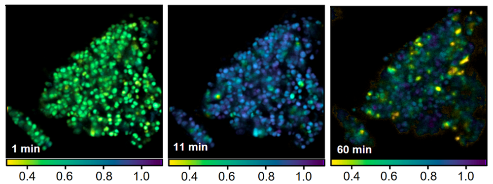
Until now, there has been insufficient data on how exactly the composition of lipofuscin changes under photodamage and how this is reflected in its “glow” signal. For the first time, Russian researchers used fluorescence lifetime imaging to track at the cellular level how lipofuscin changes during photooxidation inside pigment epithelial cells.
Experiments showed that as photooxidation progresses, not only does the composition of lipofuscin change, but also the nature of its “glow”: in particular, the fluorescence lifetime increases. This is presumably due to the fact that the original molecular components of lipofuscin are oxidized and partially broken down, while the products of their transformation have different fluorescent properties.
“What is important is that we were able to record these changes without introducing additional labels or interfering with the cell. Such measurements became possible thanks to fluorescence lifetime imaging. This is a modern microscopy method based on measuring the lifetime of excited molecular states, which allows us to obtain additional diagnostic information about tissue condition,” said Alexey Semenov, Candidate of Biological Sciences, researcher at the Laboratory of Photonic Gas Sensors at MISIS.
The scientists also studied the role of antioxidants in suppressing the phototoxic effects of lipofuscin. In the experiment, they investigated the carotenoid protein AstaP, isolated from the microalgae Coelastrella astaxanthina, which can deliver zeaxanthin, which is a natural substance that protects cells from oxidative stress. It was found that the AstaP—zeaxanthin complex slows down lipofuscin degradation: the formation of oxidized products is reduced, and complete pigment damage does not occur. Details of the study are published in the Journal of Physical Chemistry B (Q1).
“To increase the sensitivity and speed of measurements, in the next stage we plan to use quantum sensors we have developed — superconducting single-photon detectors,” said Grigory Goltsman, leading researcher at the Laboratory of Quantum Information Technologies at MISIS.
The work was carried out under the program for attracting talented young scientists under the age of 39 (postdocs) within the framework of Priority 2030 (grant No. K4-2024-3).